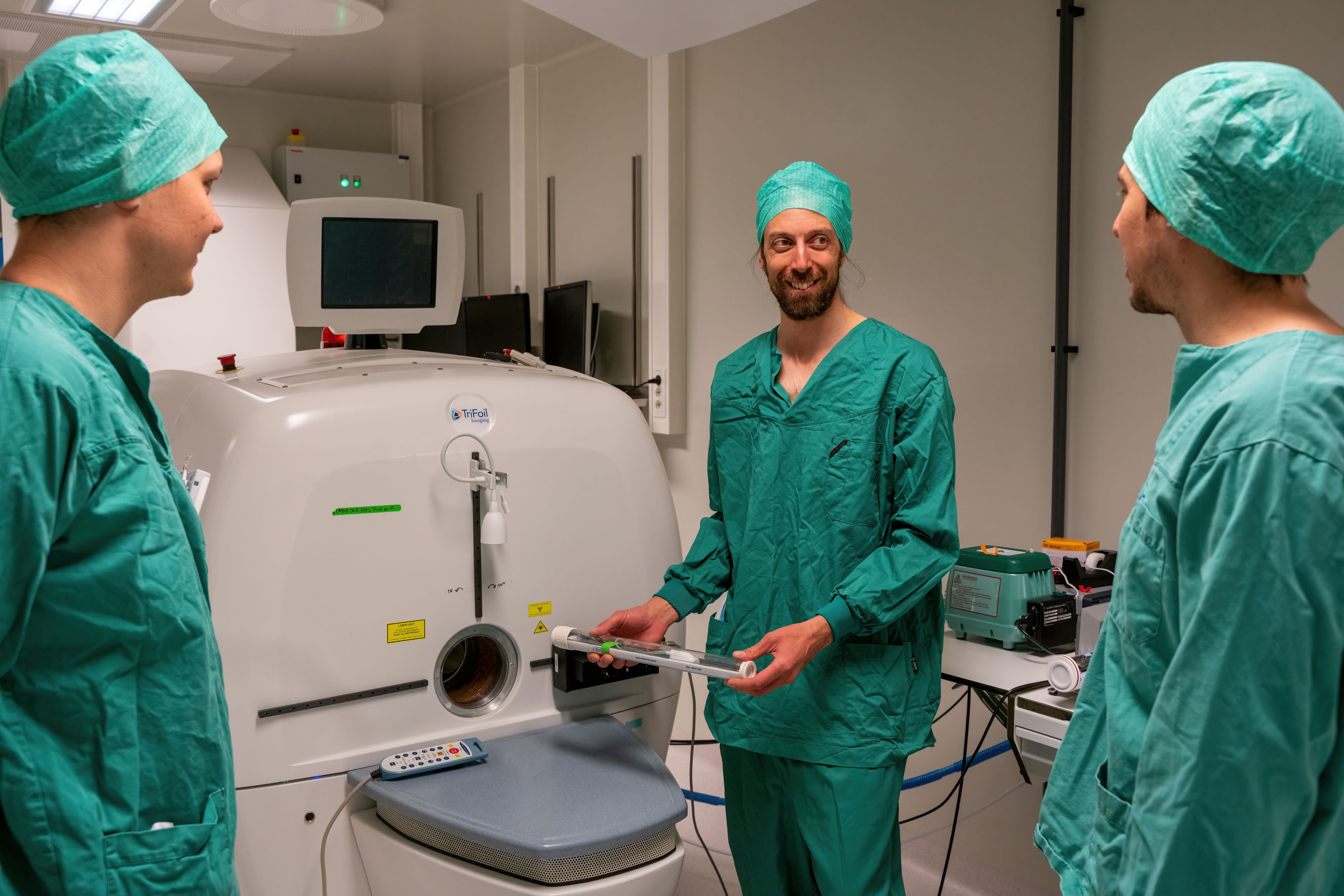



[18F]FDG Positron Emission Tomography (PET) is a widely used medical imaging technique that provides highly sensitive spatial representations of radiopharmaceuticals metabolized by tissues and cells. PET imaging is particularly valuable for identifying active tissues and detecting malignant growths, such as cancerous tumors, which appear as bright spots in the images. It is also instrumental in monitoring cancer therapies.
Traditionally, PET images are static, constructed by averaging gamma-ray counts over time after the radiotracer uptake stabilizes post-injection. While static PET is effective for many applications, its limitations become apparent in tasks like drug development, tracer design, and disease therapy research. Dynamic PET (dPET) offers an alternative by capturing radiation detections over the entire imaging duration and averaging them into smaller temporal bins. This results in a spatial time-series representation of tracer uptake, visualized through time-activity curves (TACs). These TACs reflect the temporal variability of tracer binding to proteins, free states in plasma, or metabolism. Tracer kinetic modeling, often using compartment models, estimates uptake rates between plasma, tissues, and cells. This process relies on the arterial input function (AIF), which represents plasma blood concentration.
However, obtaining an accurate AIF is challenging. The gold standard involves invasive arterial cannulation, which is complex, time-intensive, and unsuitable for long-term studies, especially in mice, as it requires terminal procedures like carotid artery sampling.
To address these challenges, non-invasive AIF estimation methods have been explored. Techniques such as population-based input functions and image-derived input functions (IDIFs) use TACs from delineated arteries or blood pools (e.g., the left ventricle). However, these methods require rigorous calibration to mitigate partial volume effects and motion artifacts, limiting their practicality. Recent advancements include machine learning-based input functions, but these approaches often depend on manual TAC delineations.In this project, we develop deep learning-based input function (DLIF) estimation methods for AIF.
Deep learning models can process entire dPET datasets and learn mappings to mimic IDIFs, including necessary calibrations. Additionally, we are advancing physics-informed deep learning models that integrate the physical dynamics of tracer uptake. These models enable voxel-wise kinetic modeling, eliminating the need for 3D segmentation steps. By combining deep learning with compartment models, we aim to provide a robust mathematical framework for describing tracer kinetics, offering insights into cellular and tissue activity at the voxel or region-of-interest level.
Read more here